Posterior Shoulder Tendon Anatomy / Anatomy - NYSORA Regional Anesthesia App / The levator scapulae muscle originates from the transverse processes of the cervical vertebra and infraspinatus muscle originates and sits in the infraspinous fossa of the scapula.
Posterior Shoulder Tendon Anatomy / Anatomy - NYSORA Regional Anesthesia App / The levator scapulae muscle originates from the transverse processes of the cervical vertebra and infraspinatus muscle originates and sits in the infraspinous fossa of the scapula.. Anatomy of the suprascapular nerve. Inserts onto navicular tuberosity and first cuneiform. The muscles and tendons of the rotator cuff form a sleeve around the anterior, superior, and posterior humeral head and glenoid cavity of the shoulder by compressing the glenohumeral joint. Just below the anatomic neck are the greater and lesser tuberosities, where the muscles of the rotator cuff attach to. Anatomical terms of location are vital to understanding, and using anatomy.
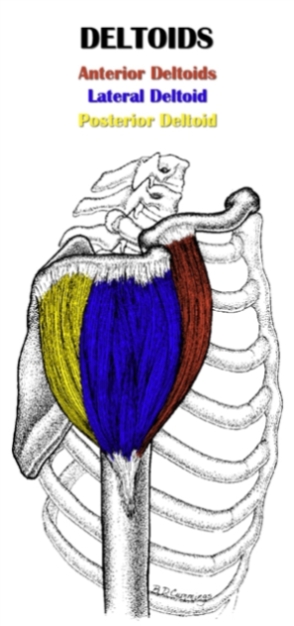
Learn about shoulder anatomy, muscles in the shoulder joints and watch anatomy of the shoulder video's presented by joi. Start studying posterior shoulder anatomy. The shoulder anatomy includes the anterior deltoid, lateral. The supraspinatus tendon and subacromial bursa). The human shoulder is made up of three bones:
Shoulder Anatomy | All About the Shoulder Muscles from www.kingofthegym.com Just below the anatomic neck are the greater and lesser tuberosities, where the muscles of the rotator cuff attach to. The supraspinatus tendon is the most commonly affected tendon in the rotator cuff. Posterior band of the ighl. Human anatomy for muscle, reproductive, and skeleton. The human shoulder is made up of three bones: One of the biceps tendons (the long head) runs in a groove (bicipital groove) that separates the two tuberosities. Right posterior belly of digastric muscle. Secondary restaint to inferior translation in the abducted shoulder.
Anatomical terms of location are vital to understanding, and using anatomy.
Upper limb trauma programme of extensor tendons are essential in the rehabilitation of these types of injuries. Upper limb, breast, posterior shoulder, lateral chest wall. Posterior — the back of the shoulder. In the shoulder, articular cartilage covers the end of the humerus and socket area of the glenoid on the scapula. Anterior graphic of the shoulder. .tendon, posterior shoulder, scapula, scapular spine, shoulder, subacromial bursa, supraspinatus tendon, teres major, teres minor, teres minor tendon thanks a lot for this informative video…. Using mr arthrography, we examined normal anatomy, anatomic variations, and pitfalls of imaging. Normal anatomy, variants and checklist. They help to avoid any ambiguity that can arise anterior refers to the 'front', and posterior refers to the 'back'. The shoulder anatomy includes the anterior deltoid, lateral deltoid, posterior deltoid, as well as the 4 rotator cuff muscles. Assoc prof craig hacking ◉ ◈ and dr jeremy jones ◉ et al. The muscles and tendons of the rotator cuff form a cover around the anterior, superior, and posterior humeral head and glenoid cavity of the shoulder by compressing. Being an undergraduate student excites me and inspires me to lean.
Posterior band of the ighl. Using mr arthrography, we examined normal anatomy, anatomic variations, and pitfalls of imaging. Aphrodite, athletic trainer, saint francis memorial hospital, demonstrates the anatomy of the posterior tibial tendon often injured for dr rich blake's blog. The muscles and tendons of the rotator cuff form a cover around the anterior, superior, and posterior humeral head and glenoid cavity of the shoulder by compressing. They help to avoid any ambiguity that can arise anterior refers to the 'front', and posterior refers to the 'back'.
Schematic representation of the right shoulder. Anterior ... from www.researchgate.net The levator scapulae muscle originates from the transverse processes of the cervical vertebra and infraspinatus muscle originates and sits in the infraspinous fossa of the scapula. Learn vocabulary, terms and more with flashcards, games and other study tools. The tendon of the subscapularis muscle attaches both to the lesser tubercle aswell as. Right posterior belly of digastric muscle. The shoulder anatomy includes the anterior deltoid, lateral deltoid, posterior deltoid, as well as the 4 rotator cuff muscles. Robin smithuis and henk jan van der woude. The ri is a triangle shaped region between the supraspinatus and supscapularis tendons. Human anatomy for muscle, reproductive, and skeleton.
Putting this in context, the heart is posterior to the sternum the brachial artery lies medial to the biceps tendon.
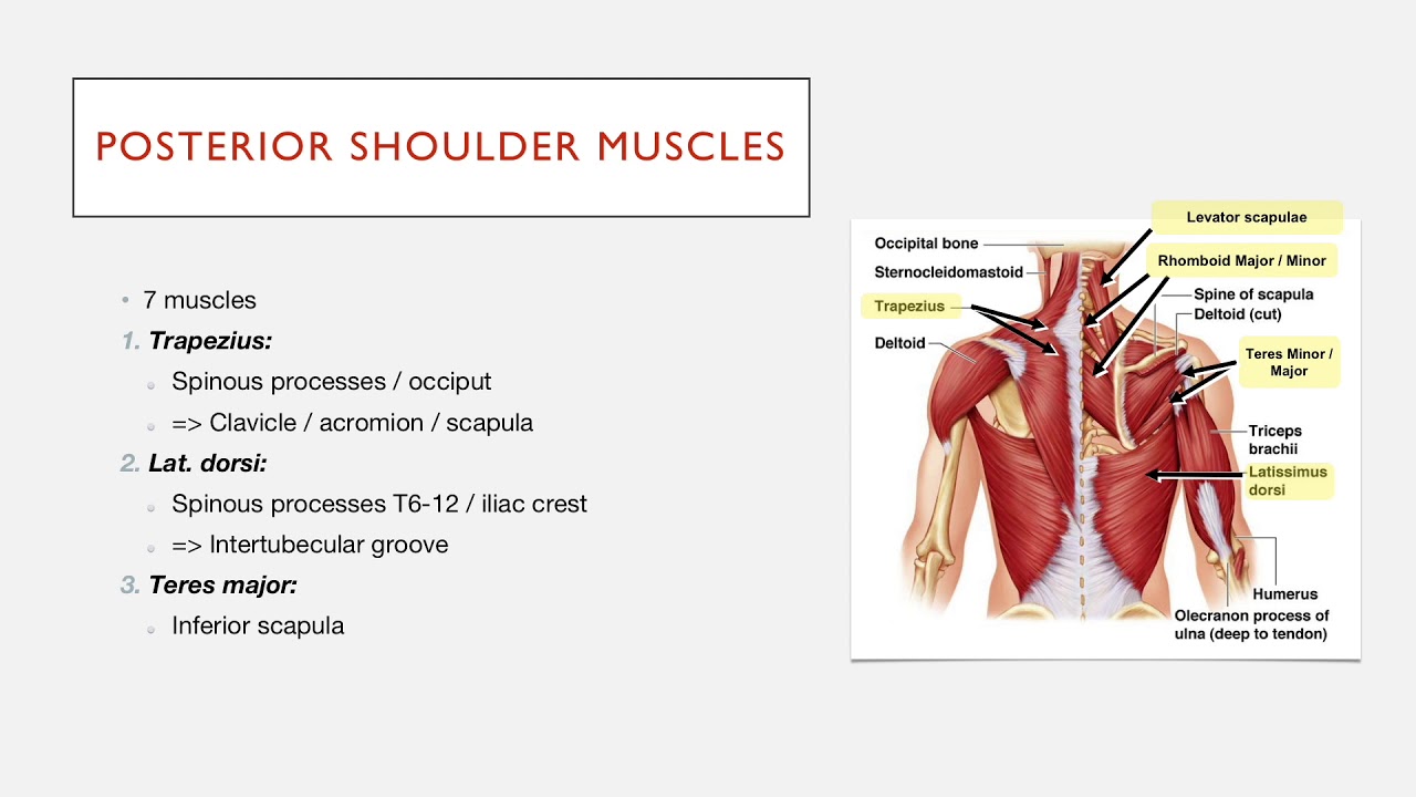
Normal anatomy, variants and checklist. Acute tears may occur when the arm is violently pushed into. An image depicting shoulder anatomy can be seen below. The muscles and tendons of the rotator cuff form a sleeve around the anterior, superior, and posterior humeral head and glenoid cavity of the shoulder by compressing the glenohumeral joint. Learn vocabulary, terms and more with flashcards, games and other study tools. Back (posterior) muscles of the shoulder. The muscles and tendons of the rotator cuff form a cover around the anterior, superior, and posterior humeral head and glenoid cavity of the shoulder by compressing. Infrspinatus tendon and teres minor. The supraspinatus tendon is the most commonly affected tendon in the rotator cuff. Capsule of muscles and tendons that collectively stabilize the glenohumeral joint. Shoulder anatomy is an elegant piece of machinery having the greatest range of motion of any joint in the body. Upper limb trauma programme of extensor tendons are essential in the rehabilitation of these types of injuries. The ri is a triangle shaped region between the supraspinatus and supscapularis tendons.
Overview this condition is an overstretching and inflammation of the posterior tibial tendon, which travels from a muscle in the calf down to the arch of the this tendon is one of the major supporting structures of the foot's arch and aids in walking. Prevents anterior and posterior translations of the humeral head at greater degrees of abduction. Anterior graphic of the shoulder. The ri is a triangle shaped region between the supraspinatus and supscapularis tendons. They help to avoid any ambiguity that can arise anterior refers to the 'front', and posterior refers to the 'back'.
Posterior Shoulder Muscles: Structure - YouTube from i.ytimg.com Assoc prof craig hacking ◉ ◈ and dr jeremy jones ◉ et al. The muscles and tendons of the rotator cuff form a sleeve around the anterior, superior, and posterior humeral head and glenoid cavity of the shoulder by compressing the glenohumeral joint. Posterior band of the ighl. The human shoulder is made up of three bones: Posterior shoulder pain is more often than not mistakenly identied as rotator cuff disease or cervical disk 9 retraction of the supraspinatus tendon in a massive rotator cuff tear leading to reduction of the acute. .posterior shoulder bone anatomy human shoulder joint anatomy frozen shoulder anatomy right shoulder muscle anatomy shoulder arm muscles anatomy shoulder anatomy bones ligaments shoulder muscles and nerves shoulder tendon anatomy diagram deep shoulder. Infraspinatus and teres minor tendon. Anterior graphic of the shoulder.
Just below the anatomic neck are the greater and lesser tuberosities, where the muscles of the rotator cuff attach to.
Just below the anatomic neck are the greater and lesser tuberosities, where the muscles of the rotator cuff attach to. Posterior shoulder instability, accelerated osteoarthritis and pos the shoulder joint is functionally and structurally complex and is composed of bone, hyaline cartilage, labrum, ligaments objective: Anatomy of the suprascapular nerve. .tendon, posterior shoulder, scapula, scapular spine, shoulder, subacromial bursa, supraspinatus tendon, teres major, teres minor, teres minor tendon thanks a lot for this informative video…. Human anatomy for muscle, reproductive, and skeleton. The muscles and tendons of the rotator cuff form a cover around the anterior, superior, and posterior humeral head and glenoid cavity of the shoulder by compressing. Being an undergraduate student excites me and inspires me to lean. Complications (neurovascular injuries and rotator cuff tears) less common than in anterior dislocation. Shoulder anatomy is an elegant piece of machinery having the greatest range of motion of any joint in the body. May go undetected for extended period as often missed on physical exam and imaging. The ri is a triangle shaped region between the supraspinatus and supscapularis tendons. Robin smithuis and henk jan van der woude. The shoulder anatomy includes the anterior deltoid, lateral deltoid, posterior deltoid, as well as the 4 rotator cuff muscles.
Related posts of wrist tendon anatomy diagrams shoulder tendon anatomy. Right posterior belly of digastric muscle.


0 Komentar